TEDx Talk: How AI Can Make Cancer Care More Equitable
Invited Talk, TEDx Nagpur Salon, Nagpur, India
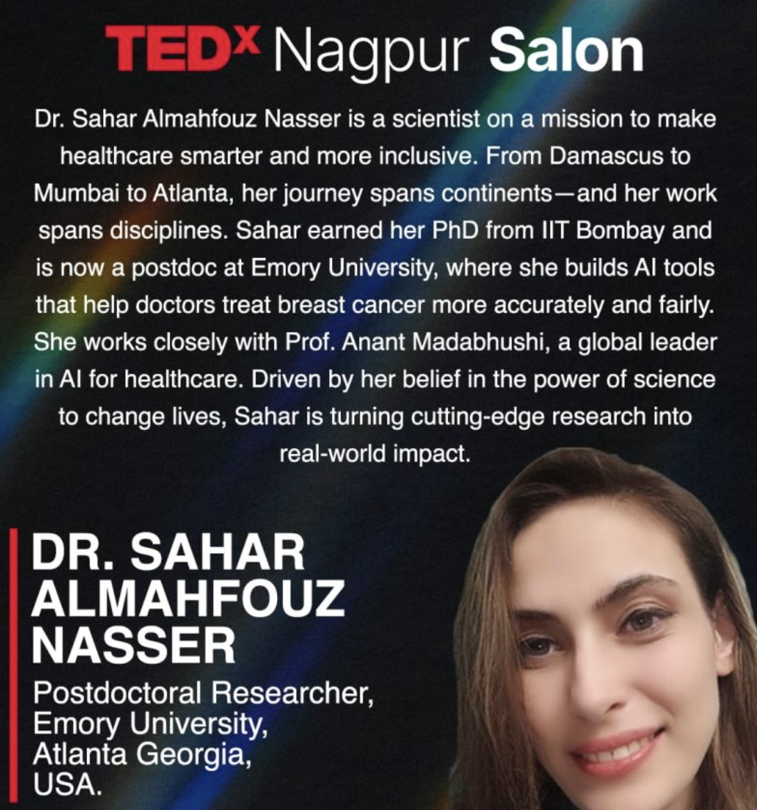
I was invited to give a TEDx Salon Talk on how AI tools are improving equitable breast cancer diagnosis and treatment.
In this talk, I shared my research journey, highlighting how computational pathology and explainable AI can bring precision diagnostics to underserved populations.
Watch the Talk
Abstract
Artificial Intelligence has the potential to democratize cancer care by reducing costs, improving diagnostic accuracy, and enabling timely interventions.
In my TEDx talk, I discussed examples from my work on developing equitable AI biomarkers, while emphasizing the importance of fairness, transparency, and accessibility in medical AI.